Ankle X Ray Report Template
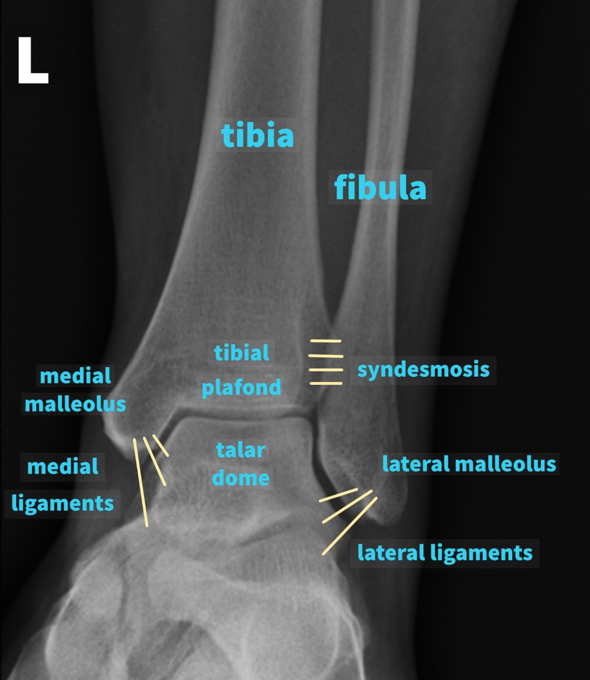
Ankle X Ray Report Template - No fracture or dislocation is identified. Web the infinity® total ankle is intended to give a patient limited mobility by reducing pain, restoring alignment and replacing the flexion and extension movement in the ankle joint. Web free download pdf word format x rays right ankle joint ap/lat/obl. Multiplane pd, pd fat sat and t1 images were obtained through the ____ ankle. Radiograph of left ankle joint obtained in anteroposterior, lateral and oblique projections. Web x rays right foot ap report template title modality x rays part right foot ap technique radiograph of right foot obtained in anteroposterior projection. No fracture or dislocation is identified. Web free download pdf word format x rays left ankle joint ap/lat. Radiographs of right ankle joint obtained in anteroposterior projection. The first two versions are blank whereas the third included examples fork every write category.
Knee X Ray Report Template (4) PROFESSIONAL TEMPLATES
Radiographs of right ankle joint obtained in anteroposterior projection. Also available other updated radiology mri, ct scan, xray, sonography, usg, mammography, pet ct,. No fracture or dislocation is identified. Web x rays left ankle joint ap/lat/obl report template. Radiographs of right ankle joint obtained in anteroposterior and lateral projection.
The appealing Chiropractic X Ray Referral Form Template Fill Online
There is no soft tissue swelling or joint effusion. No fracture or dislocation is identified. Radreport templates are intended to provide examples of best practices for diagnostic reporting. No obvious fracture is seen. Web beneath you will find acpm’s standardized radiology report template.
Ankle Xray Interpretation LaptrinhX / News
Web free download pdf word format x rays right ankle joint ap/lat/obl. Web x rays right foot ap report template title modality x rays part right foot ap technique radiograph of right foot obtained in anteroposterior projection. Radiographs of right ankle joint obtained in anteroposterior projection. The first two versions are blank whereas the third included examples fork every write.
AVN 2' to SCA sample CASE REPORT
De3d imaging was also acquired. Web beneath you will find acpm’s standardized radiology report template. There is no soft tissue swelling or joint effusion. No fracture or dislocation is identified. No obvious fracture is seen.
X Ray Report Template (11) TEMPLATES EXAMPLE Report template
Web x rays left ankle joint ap/lat/obl report template. ____ view/s of the right/left foot. Radreport templates are intended to provide examples of best practices for diagnostic reporting. Web x rays right ankle joint ap/lat report template. Multiplane pd, pd fat sat and t1 images were obtained through the ____ ankle.
61 INFO FOOT X RAY REPORT TEMPLATE PDF ZIP DOWNLOAD PRINTABLE CDR PSD
There is no soft tissue swelling or joint effusion. Web x rays left ankle joint ap/lat/obl report template. ____ view/s of the right/left ankle. Web rsna no longer publishing new templates. We provide resources to help you standardize reporting practices to enhance efficiency, demonstrate value and improve diagnostic quality.
ankle sprain in right ankle(eversion) Ankle Problems Forums Patient
No obvious fracture is seen. Web x rays right foot ap report template title modality x rays part right foot ap technique radiograph of right foot obtained in anteroposterior projection. Web on radreport.org you can: No fracture or dislocation is identified. Radiograph of left ankle joint obtained in anteroposterior, lateral and oblique projections.
Lateral radiograph demonstates a nice example of an ankle joint
No fracture or dislocation is identified. Web magnetic resonance imaging (mri). Radiographs of right ankle joint obtained in anteroposterior and lateral projection. No obvious fracture is seen. Web on radreport.org you can:
Lumbar X Ray Report Template (1) PROFESSIONAL TEMPLATES Report
Web x rays right foot ap report template title modality x rays part right foot ap technique radiograph of right foot obtained in anteroposterior projection. Also available other updated radiology mri, ct scan, xray, sonography, usg, mammography, pet ct,. Web magnetic resonance imaging (mri). Radiograph of left ankle joint obtained in anteroposterior, lateral and oblique projections. Also available other updated.
Knee X Ray Report Template (8) TEMPLATES EXAMPLE Report template
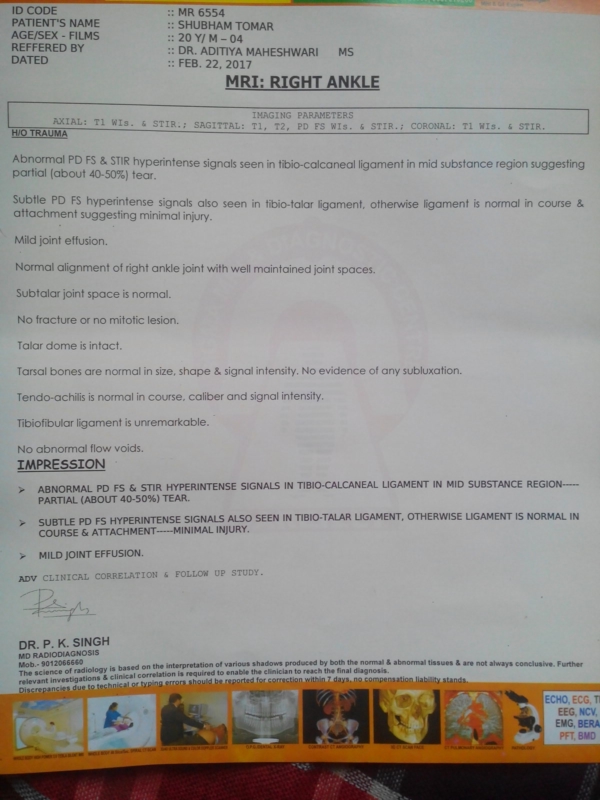
____ view/s of the right/left ankle. Multiplane pd, pd fat sat and t1 images were obtained through the ____ ankle. Web x rays right ankle joint ap/lat report template. The first two versions are blank whereas the third included examples fork every write category. Radiographs of right ankle joint obtained in anteroposterior and lateral projection.
Web on radreport.org you can: Radreport templates are intended to provide examples of best practices for diagnostic reporting. No obvious fracture is seen. Web the infinity® total ankle is intended to give a patient limited mobility by reducing pain, restoring alignment and replacing the flexion and extension movement in the ankle joint. Web x rays right ankle joint ap/lat report template. Also available other updated radiology mri, ct scan, xray, sonography, usg, mammography, pet ct,. No obvious fracture is seen. Web free download pdf word format x rays right ankle joint ap/lat/obl. We provide resources to help you standardize reporting practices to enhance efficiency, demonstrate value and improve diagnostic quality. No obvious fracture is seen. Also available other updated radiology mri, ct scan, xray, sonography, usg, mammography, pet. ____ view/s of the right/left ankle. No fracture or dislocation is identified. Web magnetic resonance imaging (mri). Web x rays left ankle joint ap/lat/obl report template. Web beneath you will find acpm’s standardized radiology report template. Web x rays right ankle ap report template. ____ view/s of the right/left foot. Web x rays right foot ap report template title modality x rays part right foot ap technique radiograph of right foot obtained in anteroposterior projection. No fracture or dislocation is identified.


